Tl/DR:
Our cells run on precision, and their balance depends on tiny molecular anchors i.e. membrane proteins that keep proteins in place.GG support these anchors, securing proteins to cell membranes where they manage energy, signaling, and repair.
Ever wondered how cells stay so organized?
Well, this order is maintained by membrane anchoring mechanisms which ensures proteins remain exactly where they need to be. These molecular anchors secure enzymes and signaling proteins to the cell membrane, allowing efficient energy production, clear communication, and quick repair.
Without proper anchoring, cellular coordination might fail, causing a decrease in energy levels, coordination, and resilience. Therefore, membrane anchoring is essential for healthy cellular function and aging.
To understand how anchoring works, you first need to understand what cell membranes are and why they are the central platform for cellular control. Let’s discuss in next section.
What Is a Cell Membrane?
The cell membrane, also known as the plasma membrane, is a flexible, living barrier that surrounds every cell and maintains its internal environment. It separates the intracellular (inside) and extracellular (outside) fluids while allowing controlled exchange between them.(1)
Structure and Composition of cell membrane
- The membrane is built from a phospholipid bilayer; two layers of lipid molecules arranged tail to tail.
- Each phospholipid has:
- A hydrophilic (water-loving) head made of phosphate.
- Two hydrophobic (water-repelling) tails made of fatty acids.
- This arrangement creates an amphipathic structure (hydrophilic polar part-water loving, hydrophobic nonpolar part-water shy) that makes the membrane selectively permeable(1)
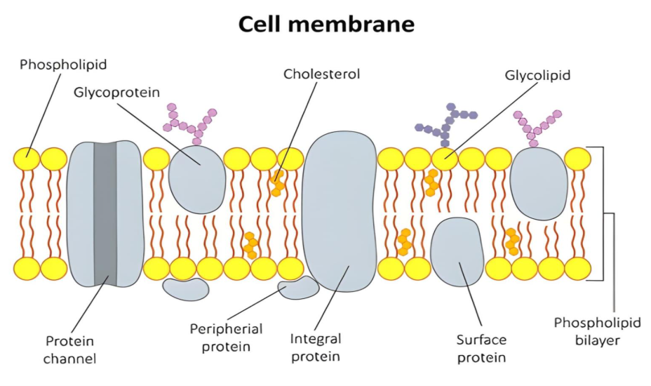
Inside this membrane, a family of molecules called membrane proteins work to move substances, transmit signals, and support the cell’s structure.
Many proteins need to anchor themselves to cell membrane and that’s exactly where Geranylgeraniol (GG) steps in. Interestingly, this small molecule plays a much bigger role than it seems. (2) Let’s unfold in the upcoming section.
Ever wondered how proteins secure cellular stability? Let’s explore how these remarkable proteins play a vital role in maintaining structural order and ensuring smooth communication within cells.
Gatekeepers of Life: Understanding Membrane Proteins
- Membrane proteins regulate cellular traffic by deciding what goes in, what stays out, and which signals need to be transmitted.
- They sieve essential molecules and nutrients while blocking harmful substances.
- They relay signals between cells, ensuring tissues and organs work together in harmony.
- They support energy flow, keep the cell stable and organized, so everything works in the right place at the right time.
Membrane proteins are truly the gatekeepers of life as they control access, protect the cell, and direct the flow of energy and communication that keeps every cell alive and functioning. Without them, the cell’s defenses would crumble, and the entire system of life from muscle strength to hormonal balance would lose its order.
Now that we understand their importance, let’s look at the main types of membrane proteins and what each of them does inside the cell.
Types of membrane proteins
Depending on how they interact with the membrane, they are divided into three main types:
1. Integral (Intrinsic) Proteins
- Go deep into or across the membrane.
- Acts as channels and receptors to move materials and receive messages like Ion channels and ATP synthase.
- GG helps maintain the lipid environment that keeps these proteins active and stable (2)
- Glycoproteins: Proteins with carbohydrate chains extending into the extracellular space. These act as identification tags, helping cells recognize each other and forming part of a protective layer called the glycocalyx (1)
Did You Know?
Cells wear a sugar coat called the glycocalyx!
This soft, protective layer helps your cells recognize each other, communicate, and even defend against stress.
It is also where many anchored proteins attach, using GG as a natural “molecular glue” to stay in place and keep your cell signals strong.
Peripheral (Extrinsic) Proteins
- Loosely attached to the membrane surface or other proteins.
- Support enzyme functions and signaling like: Cytochrome c, G proteins.
- Many peripheral proteins require lipid modification to stay attached, a process supported by GG-derived lipids (1,2)
Cytochrome C: The Dual-Role Molecule
Tucked inside mitochondria; cytochrome C helps in transferring electrons during energy production (ATP). It’s like a courier that keeps your cell’s power supply running smoothly.
Lipid-Anchored Proteins
- Linked to the membrane through lipid molecules such as geranylgeranyl, farnesyl, or palmitoyl groups.
- These lipid anchors are built from the mevalonate pathway, where Geranylgeraniol (GG) serves as a key intermediate
- Example: Ras, Rho, and Rab GTPases — proteins crucial for cell signaling and transport.(2,3)
Now that we know the types of membrane proteins, let’s see how they actually anchor to membrane.
Did You Know?
The mevalonate pathway secretly works as your cell’s anchoring factory; making lipid tags that help proteins stick to membranes. Without it, those vital proteins would wander freely, unable to reach their true destination.
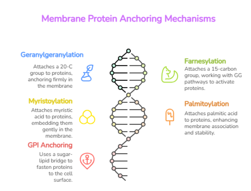
Membrane Protein Anchoring Mechanism
Even though the membrane is fluid, proteins need to stay anchored in specific spots to function correctly. This is achieved through lipid-based anchoring mechanisms, many of which rely directly on GG for building the anchor itself (3,4).
Main Anchoring Mechanisms
Geranylgeranylation
- The cell uses GG to create a 20-carbon geranylgeranyl group, which attaches to the protein’s tail.
- This bond allows signaling proteins (like Ras, Rho, and Rab) to anchor firmly into the cell membrane
- These anchored proteins manage cell growth, energy balance, and communication.(5)
Farnesylation
- A related process where a 15-carbon farnesyl group is attached instead of a geranylgeranyl group.
- Works together with GG-related pathways to keep signaling proteins active(5)
Did You Know?
Your cells use tiny “tails” to help certain proteins stick to their membranes — a bit like plug wires connecting gadgets to power!
These tails can be short (farnesyl) or long (geranylgeranyl), both made through the mevalonate pathway.
The longer tail (geranylgeranylation ) helps the protein stay firmly anchored, especially in light-sensing cells of the eye.
Myristoylation
- Myristoylation is cell’s first step in which a fatty acid tag “myristic acid “attaches to the protein’s first glycine, giving it a subtle hydrophobic edge.
- The enzyme N-myristoyltransferase (NMT) transfers this lipid from myristoyl-CoA, marking the start of the anchoring journey.
- This modification allows proteins to get embedded gently into the inner cell membrane, where vital signaling begins. Thus, supporting essential functions like signal transmission, enzyme activation, and viral assembly.
Both myristoylation and geranylgeranylation are lipid-anchoring mechanisms that help proteins stay attached to membranes, and both trace their origin to the mevalonate pathway, your cell “lipid factory”.(6)
Supplementing with GG helps restore harmony in the mevalonate pathway, supporting smooth protein signaling, balanced energy flow, and optimal membrane function acting as the core of cellular vitality.
4.Palmitoylation
- A lipid-based post-translational modification where a fatty acid, palmitic acid (C16), is covalently attached to cysteine residues of proteins via a thioester bond.
- Main Function is to Enhances protein-membrane association, influencing localization, stability, secretion, and signaling.(7)
Myristoylation often partners with palmitoylation or prenylation (via the mevalonate pathway) to stabilize membrane attachment.
5.GPI Anchoring (Glycosylphosphatidylinositol)
- Glycosylphosphatidylinositol (GPI) acts like a molecular hook, fastening many proteins to the cell surface through a sugar–lipid bridge.
- Found widely in humans and other eukaryotes, GPI-anchored proteins play vital roles as receptors, enzymes, and transporters, keeping cell communication and structure in perfect synchronization.
- Although GG doesn’t form this anchor directly, it supports the lipid synthesis necessary for the process
These microscopic lipid imprints determine a protein’s place, function, and lifespan proving that precision at the molecular level shapes the rhythm of cellular life.
Connecting the dots: How GG drives protein anchoring
Protein anchoring depends heavily on lipid attachment processes such as prenylation, and GG serves as a critical lipid donor in this process. Before exploring how it works, let’s first understand what GG is?
What is GG?
GG is a naturally occurring isoprenoid (a type of lipid molecule). Inside cells, it is converted into geranylgeranyl pyrophosphate (GGPP), which is used in a process called protein geranylgeranylation.
How GG Helps Proteins Attach to Membranes?
Many membrane-associated proteins can’t naturally stick to the cell membrane as they need a hydrophobic “anchor” added to them so they can insert into or associate with the lipid bilayer. Here’s where GG comes in:
1. Activation
GG is converted to geranylgeranyl pyrophosphate (GGPP) in the cell.
2. Attachment (Geranylgeranylation)
Specialized enzymes called geranylgeranyl transferases (GGTases) transfer the geranylgeranyl group from GGPP onto specific proteins usually at a cysteine residue near the protein’s C-terminus (end of the protein).
3. Anchoring to the Membrane
The geranylgeranyl group is hydrophobic, so once attached, it embeds itself into the lipid bilayer of the cell membrane. This tethers the protein to the membrane, allowing it to:
- Interact with other membrane proteins,
- Transmit signals,
- Move materials, or
- Help maintain cell structure.(5,8)
| Study (Author, Year) | Study Design / Model | Key Findings | Relevance to GG Anchoring Mechanism |
| Zhang & Casey, 1996 (Annu Rev Biochem)(9) | Review of molecular mechanisms of protein prenylation and enzyme functions (GGTase I/II). | Identified geranylgeranylation as a post-translational modification that attaches geranylgeranyl groups from GGPP to proteins (like Ras, Rho, Rab), essential for their membrane localization. | Established the core biochemical process showing how GG → GGPP → geranylgeranylation enables protein anchoring. |
| Casey & Seabra, 1996 (J Biol Chem)(10) | Biochemical analysis of prenyltransferase enzymes and lipid donor pathways. | Explained how GG is metabolized to GGPP, the active substrate for geranylgeranyl transferases (GGTases). | Provided evidence that GG serves as a metabolic precursor for protein anchoring through enzymatic transfer. |
| Berndt et al., 2011 (Nat Rev Cancer)(11) | Review of Ras and Rho GTPases in cancer cell signaling and prenylation inhibition studies. | Blocking geranylgeranylation causes Ras/Rho proteins to mislocalize from membranes to the cytosol, shutting down key signaling pathways. | Demonstrated the functional consequence of GG deficiency — loss of protein anchoring and disrupted signaling. |
| Ho et al., 2016 (Biochem Biophys Res Commun)(12) | In vitro study on testis-derived I-10 cells investigating GG’s role in cellular signaling. | GG supplementation increased testosterone synthesis and enhanced cAMP/PKA signaling, showing GG’s ability to support lipid-mediated pathways. | Indirectly confirmed GG’s biological activity in restoring proper signaling via lipid modifications. |
Evidence Supporting the Role of Geranylgeraniol (GG) in Protein Anchoring and Cellular Function
Did You Know?
Small proteins like Ras and Rho need GG to “stick” to cell membranes. Without GG, they float freely in the cytosol, and vital cell signaling shuts down.
Also Read: Geranylgeraniol Explained: Benefits, Side Effects & Science Behind It
Summary
One fascinating aspect of this biological choreography is how membrane proteins stay precisely where they need to be. The mechanism of membrane protein anchoring explains how these essential molecules attach to the lipid bilayer and maintain the functional architecture of the cell. GG acts as a foundation molecule for anchoring many essential proteins.
Without enough GG, key cellular proteins lose their attachment, disrupting:
- Energy production (mitochondrial signaling)
- Cell growth control
- Neurotransmission and immune balance
Low GG levels (as seen during aging or statin use) can lead to poor protein anchoring and weak cellular communication.
By restoring GG levels, it’s possible to support proper protein attachment, improve signaling, and maintain cellular vitality, making GG an emerging focus in biomedical and wellness research.
Key Takeaways
- The cell membrane protects and organizes all cellular activities.
- Proteins embedded in or attached to it control communication, transport, and energy flow.
- Geranylgeraniol (GG) provides the lipid “anchors” that keep these proteins stable and functional.
- Maintaining GG levels ensures strong cell signaling, energy balance, and healthy cellular function.
FAQ’s
GG provides lipid tails (geranylgeranyl groups) that act like “molecular hooks,” helping proteins stay attached to membranes.
Low GG levels disrupt cell signaling and energy flow, which may cause muscle fatigue or slower recovery.
GG is synthesized through the mevalonate pathway, the same route that produces cholesterol and CoQ10.
Yes. GG supports mitochondrial energy, muscle function, and protein anchoring.
Yes. GG is naturally made in your cells and found in foods like olive oil and annatto. However, supplementation with GG adds value to your health.
References
- Anamourlis C. The cell membrane. South Afr J Anaesth Analg. 2020;26(6 Suppl 3):S2–S5. doi:10.36303/SAJAA.2020.26.6.S3.2527.
- asas J, Ibarguren M, Álvarez R, Terés S. G protein–membrane interactions II: Effect of G protein-linked lipids on membrane structure and G protein–membrane interactions. Biochim Biophys Acta Biomembr. 2017;1859(9 Pt B):1523-1535.. doi:10.1016/j.bbamem.2017.05.018
- Casas J, Ibarguren M, Álvarez R, Terés S, Lladó V, Piotto SP, Concilio S, Busquets X, López DJ, Escribá PV. G protein–membrane interactions II: Effect of G protein-linked lipids on membrane structure and G protein–membrane interactions. Biochim Biophys Acta Biomembr. 2017;1859(9 Pt B):1523-1535. doi:10.1016/j.bbamem.2017.05.018
- Zhang FL, Casey PJ. Protein prenylation: molecular mechanisms and functional consequences. Annu Rev Biochem. 1996;65:241–269. doi:10.1146/annurev.bi.65.070196.001325
- Kassai H, Fukada Y. Farnesylation versus geranylgeranylation in G-protein-mediated light signaling. J Biol Chem. 2011;286(11):8687–8696. doi:10.1074/jbc.M110.203216
- Cao W, Sumikoshi K, Nakamura S, Terada T, Shimizu K. Prediction of N-myristoylation modification of proteins by SVM. Bioinformation. 2011;6(2):62-63. doi:10.6026/97320630006062
- Li W, Shen J, Zhuang A, Wang R, Li Q, Rabata A, Zhang Y, Cao D. Palmitoylation: an emerging therapeutic target bridging physiology and disease. Cell Mol Life Sci. 2023;80(1):25. doi:10.1007/s00018-022-04671-7
- Yuan Y, Li P, Li J, Zhao Q, Chang Y, He X. Protein lipidation in health and disease: molecular basis, physiological function and pathological implication. Signal Transduct Target Ther. 2024;9:60. doi:10.1038/s41392-024-01759-7.
- Zhang FL, Casey PJ. Protein prenylation: molecular mechanisms and functional consequences. Annu Rev Biochem. 1996;65:241-269. doi:10.1146/annurev.bi.65.070196.001325
- Casey PJ, Seabra MC. Protein prenyltransferases. J Biol Chem. 1996;271(10):5289-5292. doi:10.1074/jbc.271.10.5289
- Berndt N, Hamilton AD, Sebti SM. Targeting protein prenylation for cancer therapy. Nat Rev Cancer. 2011;11(11):775-791. doi:10.1038/nrc3151
- Ho TT, Murakami M, Islam S, et al. Geranylgeraniol enhances testosterone production via cAMP/PKA signaling in I-10 cells. Biochem Biophys Res Commun. 2016;474(3):521-526. doi:10.1016/j.bbrc.2016.04.130