Tag: Geranylgeranoil

  • Stronger From the Inside: The GG Way of Oxidative Stress Management

    Stronger From the Inside: The GG Way of Oxidative Stress Management

    Tl/DR:

    Your strongest antioxidant protection comes from within. GG supports CoQ10 and mitochondrial health, making your cells more resilient against oxidative stress.

    What if the air you breathe, the processed foods you eat, and hours of sitting were quietly damaging your cells? Modern living overloads your body with oxidative stress, thereby weakening energy, focus, metabolism, and accelerating aging. Your cells fight back with antioxidants, but sometimes, it’s not about taking only Vitamin C or E.

    The real key to protection is boosting your body’s own antioxidant engine, and that’s where Geranylgeraniol (GG) plays a crucial role. It supports the production of CoQ10 and other cellular antioxidants, helping your cells stay energized, resilient, and protected against oxidative damage.

    What is Oxidative Stress?

    • Oxidative stress happens when your body produces more free radicals than it can neutralize.
    • Free radicals are unstable molecules generated from normal metabolism but their levels spike with pollution, smoking, processed foods, chronic stress, and certain medications.(1)
    • Under normal conditions, your body constantly produces free radicals (from breathing, metabolism, immune responses) and neutralizes them using antioxidants like glutathione, vitamin C, vitamin E, and antioxidant enzymes (SOD, catalase)
    • When free radical production rises too high (pollution, smoking, inflammation, certain drugs) or antioxidant defenses drop too low (poor diet, illness, aging), this balance tips. That imbalance is what we call oxidative stress.(1)
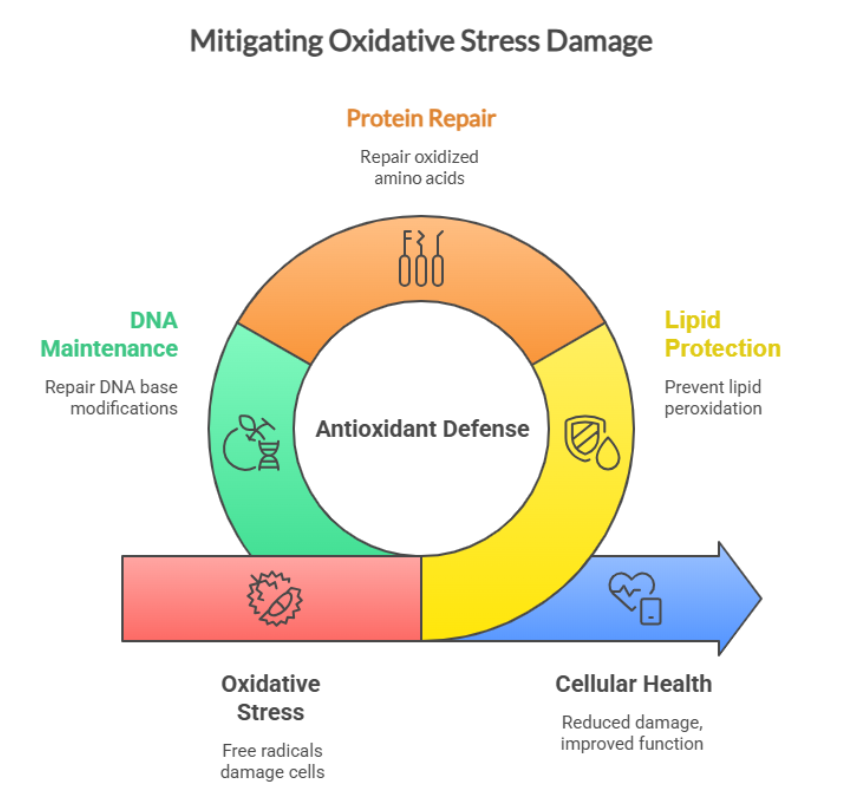
    • When these reactive molecules build up, they begin attacking essential cellular components:
      • Lipids → causing membrane damage and impaired cell signaling
      • Proteins → altering structure and function of enzymes and tissues
      • DNA → leading to mutations, accelerated aging, and increased disease risk(2)
    Mitigating Oxidative Stress Damage

    What happens when antioxidant defense falls behind?

    When lipid and protein accumulate, and DNA damage occurs faster than the body can repair it, oxidative stress causes fatigue, premature aging, cardiovascular disease, diabetes, neurodegeneration, kidney disease and chronic inflammation.

    But here’s the reassuring part: your body has its own antioxidant machinery designed to handle this stress. Let’s discuss this further in the next section.

    How Your Body’s Built-In Antioxidant System Protects You

    Your body is equipped with a highly sophisticated internal antioxidant network that works around the clock to neutralize free radicals, repair damage, and keep your cells functioning at their best. These include Glutathione, antioxidant enzymes (catalase, glutathione peroxidase), and Coenzyme Q10 (CoQ10).

    • At the center of this defense is glutathione (GSH), often called the body’s “master antioxidant.”
      • Glutathione directly neutralizes reactive oxygen species (ROS) and detoxifies harmful byproducts before they can injure your cells.
      • When glutathione levels drop (aging, chronic stress, or inflammation), the body becomes far more vulnerable to oxidative damage and metabolic slowdown.
    • Working alongside glutathione are powerful antioxidant enzymes, working day and night as quiet, tireless warriors.
    Superoxide dismutase (SOD) converts highly reactive superoxide radicals into hydrogen peroxide, while catalase and glutathione peroxidase (GPx) break that hydrogen peroxide down into harmless water and oxygen.
    • Together, these enzymes protect your mitochondria, defend DNA, and reduce inflammation at the cellular level.
    • Another essential player is CoQ10; the antioxidant embedded deep within mitochondrial membranes.
      • CoQ10 supports in ATP energy production
      • It also stabilizes mitochondrial structure and
      • It prevents lipid peroxidation, making it especially important for the heart, brain, and muscle health.
    Strong CoQ10 levels = stronger antioxidant protection, better energy, and healthier aging.
    Internal Antioxidant Network 

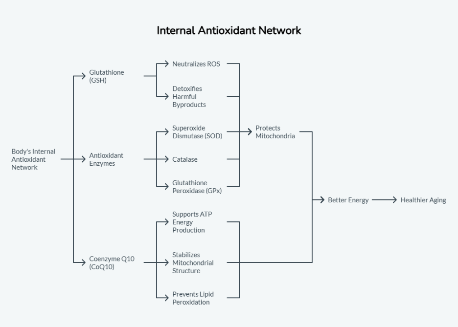
    All these components work as your internal “antioxidant security team,” keeping oxidative stress under control so you can stay energized, mentally sharp, and biologically resilient.

    And here’s the key connection: Instead of acting as a direct antioxidant, geranylgeraniol (GG) helps your body strengthen this entire built-in defense system. Let’s understand this in next section.

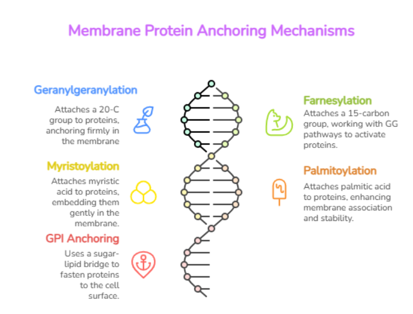
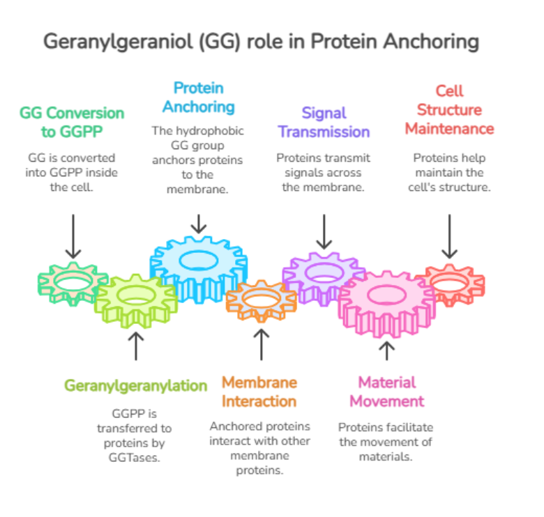
    Geranylgeraniol (GG) is an isoprenoid naturally produced in the mevalonate pathway, the biochemical highway that generates cholesterol, steroid hormones, vitamin D, and CoQ10. GG plays three major roles

    • It fuels CoQ10 biosynthesis
    • Supports mitochondrial function
    • Maintains cellular structure through protein prenylation(4)

    Read more: A comprehensive guide to GG supplement

    How GG Supports Antioxidant Capacity

    1. GG Helps Your Body Make More CoQ10

    • CoQ10 is one of the most powerful antioxidants in human physiology, but the body relies on the mevalonate pathway to produce it.
    • GG is a crucial intermediate in this pathway. Without enough GG, CoQ10 production slows down, leaving mitochondria more vulnerable to oxidative damage. This explains why low-GG states (aging, statins) often coincide with fatigue and muscle weakness.(5)

    Reduced GG → Reduced CoQ10 → weakened antioxidant defense.

    Read more: CoQ10 Benefits: What is Coenzyme Q10 Used For?

    2. GG Improves Mitochondrial Quality 

    • Healthy mitochondria produce fewer free radicals.
    • Studies such as Jiwan et al. (2022, In Vivo) show that GG supplementation improved mitochondrial enzyme function and reduced oxidative stress markers in diabetic rats leading to healthier muscle tissue. (6)
    • Better-functioning mitochondria are more efficient at energy production and generate far less oxidative “waste.”

    3. GG Reduces Inflammation-Driven Oxidative Stress 

    • Chronic inflammation is one of the biggest drivers of oxidative stress.
    • Research done by Chung et al. (2021, Nutrition Research) demonstrated that GG supplementation lowered inflammatory cytokines like IL-6 and MCP-1 in obese mice.
    • By quieting inflammation, GG helps reduce the free radical load that inflammation normally triggers.(7)
    Study Year Design N Model Dose Duration Key Results 
    Shen C-L et al. Effect of Dietary GG and Green Tea Polyphenols on Inflammation and Oxidative Stress in Obese Mice(8) 2023 Animal study Not reported Obese mice on high-fat diet 400 mg/kg diet 12 weeks GG reduced pro-inflammation and oxidative stress by inhibiting NF-KB activation; improved overall redox balance 
    Meister M et al. Dietary Geranylgeraniol and Statins May Act in Synergy to Improve Metabolic Health(9) 2022 Animal study Not reported Obese mice treated with statins ± GG 400 mg/kg diet 10 weeks GG improved mitochondrial function and  reduced oxidative stress in statin-treated mice; restored redox homeostasis via the mevalonate pathway 

    Evidence table: Recent Studies on GG and Oxidative Stress Reduction

    Now, that we have seen how GG backs up your antioxidant system, you might wonder, how is it different from usual antioxidants we take? Let’s find out.

    How GG Differs from Traditional Antioxidants

    Most antioxidants we consume (vitamin C, vitamin E, polyphenols) work by directly neutralizing free radicals. GG works differently. It supports the internal pathways that power antioxidant production and mitochondrial efficiency. By supporting upstream pathways, GG enhances the body’s own long-lasting antioxidant machinery.(10)

    Direct antioxidants are firefighters. GG helps in preventing fire in the first place.

    Conclusion

    Oxidative stress is universal and to counteract , your body has powerful built-in defenses. By supporting the mevalonate pathway and CoQ10 production, GG strengthens your antioxidant foundation from the inside out. Instead of working like a typical antioxidant, GG fuels the underlying system that keeps your cells protected, energized, and more resilient against daily oxidative challenges.

    For individuals seeking deeper cellular support, GG is emerging as a promising, science-backed approach to oxidative stress management.

    Key Takeaways

    Oxidative stress occurs when free radicals exceed your body’s antioxidant capacity, leading to cellular damage, fatigue, and accelerated aging.

    Your body has a built-in antioxidant defense system(glutathione, CoQ10, SOD, catalase, and GPx)that protects cells around the clock.

    CoQ10 supports energy production and prevents lipid peroxidation, especially in heart and muscle tissues.

    Geranylgeraniol (GG) fuels the mevalonate pathway, enabling CoQ10 biosynthesis and cellular repair processes.

    GG strengthens the antioxidant system from the inside out, instead of acting like a direct antioxidant (e.g., vitamin C or E).

    FAQ’S 

    Q1. What makes GG different from regular antioxidants? 

    Unlike vitamin C or E, GG doesn’t neutralize free radicals directly. Instead, it supports CoQ10 production and mitochondrial function thereby strengthening the body’s internal antioxidant defense system.

    Q2. How does GG help reduce oxidative stress?

    GG boosts CoQ10 biosynthesis, improves mitochondrial efficiency, and lowers inflammation-driven ROS production. These upstream effects reduce oxidative damage at the cellular level. You can read more on this

    Q3. Is GG helpful for people taking statins?

    Statins reduce GG and CoQ10 through mevalonate pathway inhibition. Supplementing GG may help maintain CoQ10 levels and support muscle health. Always consult your healthcare provider.

    Q4. Does GG act as an antioxidant?

    Not directly. GG supports the pathways that produce antioxidants like CoQ10 and glutathione, making it a potent upstream antioxidant enhancer.

    Q5. Who benefits most from GG supplementation?

    Adults over 40, statin users, athletes, people dealing with chronic stress, inflammation, pollution exposure, or low energy levels.

    References 

    1. Pizzino G, Irrera N, Cucinotta M, et al. Oxidative stress: harms and benefits for human health. Oxid Med Cell Longev. 2017;2017:8416763. doi:https://doi.org/10.1155/2017/8416763 
    1. Valko M, Leibfritz D, Moncol J, Cronin MTD, Mazur M, Telser J. Free radicals and antioxidants in normal physiological functions and human disease. Int J Biochem Cell Biol. 2007;39(1):44-84. doi:10.1016/j.biocel.2006.07.001. 
    1. Yang K, Cao F, Xue Y, Tao L, Zhu Y. Three classes of antioxidant defense systems and the development of postmenopausal osteoporosis. Front Physiol. 2022;13:840293. doi:10.3389/fphys.2022.840293. 
    1. MuseChem. The Science of Geranylgeraniol: Why It Matters for Your Health. MuseChem Blog. Published March 13, 2025. 
    1. Tan B, Chin KY. Potential role of geranylgeraniol in managing statin-associated muscle symptoms. Front Physiol. 2023;14:1246589. doi:10.3389/fphys.2023.1246589 
    1. Jiwan NC, et al. Geranylgeraniol supplementation mitigates soleus muscle mitochondrial dysfunction in diabetic rats. Int J Oncol Res. 2022;36(6):2638-2646. 
    1. Chung E, Elmassry MM, Cao JJ, Kaur G, Dufour JM, Hamood AN, Shen C-L. Beneficial effect of dietary geranylgeraniol on glucose homeostasis and bone microstructure in obese mice is associated with suppression of proinflammation and modification of gut microbiome. Nutr Res. 2021;93:27-37. doi:10.1016/j.nutres.2021.07.001 
    1. Shen C-L, Elmassry MM, Cao JJ, et al. Beneficial effect of dietary geranylgeraniol on glucose homeostasis and bone microstructure in obese mice is associated with suppression of proinflammation and modification of gut microbiome. Nutr Res. 2021;93:27-37. doi:10.1016/j.nutres.2021.07.001 
    1. Meister M, Shen C-L, Feresin R. Dietary geranylgeraniol and statins may act synergistically to mitigate oxidative stress in obese mice. Int J Environ Res Public Health. 2022;19(11):6639. doi:10.3390/ijerph19116639. PMCID: PMC9193629
    1. Hashim OA, Numan IT, Mohammed NH. The protective effect of coadministration of coenzyme Q10 and vitamin E on myopathy induced by simvastatin in rats. Toxicol Rep. 2025;14:101942. doi:10.1016/j.toxrep.2025.101942. PMID: 40612652. PMCID: PMC12223430. 
  • Cell Cycle Regulation and Growth Factor Signaling

    Cell Cycle Regulation and Growth Factor Signaling

    Tl/DR:

    The cell cycle runs your body’s repair and renewal system. Growth factors guide it, checkpoints control it, and Geranylgeraniol (GG) maintains stable signaling and energy flow, so cells divide safely and efficiently.

    Have you ever wondered, how your skin heals after a scratch, how your gut lining renews itself, and how your immune system stays ready for action.

    Feels nothing short of magic, right? But that magic “the cell cycle” is your body’s most meticulous program where millions of cells are making life or death decisions: to grow, to pause, to divide or to repair. Even though we rarely think about it, this cycle is the master engine behind tissue growth, repair, and overall vitality.(1)

    These decisions depend on two things working in perfect sync:

    • Cell cycle regulation, which determines when a cell should divide
    • Growth factor signaling, which provides the external cues that tell a cell what to do

    Uncontrolled or poorly timed cell division can lead to DNA damage, chronic inflammation, or even cancer.(2)

    Behind these signals sits the mevalonate pathway, best known for producing cholesterol but just as critical for producing the isoprenoids that anchor key signaling proteins.

    One of those isoprenoids is Geranylgeraniol (GG), the precursor to GGPP (Geranylgeranylpyrophosphate), which is required for proper functioning of the small GTPases that keep growth factor signaling precise.GG helps support small GTPases, which act as little molecular “switches” that pass growth factor signals inside the cell. 3)When these switches work smoothly, your cells stay in synchronization.

    Let’s explore this in detail in the next sections.

    What Is the Cell Cycle?

    The cell cycle is the sequence of events by which a cell grows, replicates its DNA, and divides into two new daughter cells.

    It is divided into two major stages:

    1. Interphase (G1, S, G2)

    This is the cell preparation stage where it grows, checks its environment, and copies its DNA. It further consists, of G1, S, and G2 phases.

    G1 phase: Cell grows, builds energy, and produces proteins required for DNA replication.

    S phase: DNA synthesis occurs; Cyclin A & Cyclin E activate CDK2 to drive replication.

    G2 phase: Cell performs final checks, ensures DNA is error-free, and prepares for mitosis.

    Interphase = Growth + DNA replication + quality checks
    1. Mitotic Phase (M phase)

    This is where the actual division happens. It includes:

    • Mitosis: Division of the nucleus. It can be further subdivided into various types:
      • Prophase, Prometaphase, Metaphase, Anaphase, Telophase
    • Cytokinesis: Division of the cytoplasm → two identical daughter cells{4)
    M phase = Nuclear division + Cytoplasmic division

    Each phase depends on the proper completion of the previous phase, keeping the cycle tightly controlled. They are essential for growth, immunity, tissue maintenance, and reproduction.

    Main Phase Sub-Phase Key Events Checkpoint / Control 
    Interphase G1 (Gap 1) • Cell grows and accumulates energy  
    • Produces proteins needed for DNA replication 
    G1/S checkpoint: Ensures sufficient nutrients, organelles, and no DNA damage 
    Interphase S (Synthesis) • DNA is duplicated  
    • Cyclin A & Cyclin E activate CDK2 to drive DNA synthesis 
    S-phase checkpoint: ATM/ATR → CHK1/CHK2 monitor DNA structure and replication accuracy 
    Interphase G2 (Gap 2) • Final preparation for cell division  
    • Confirms DNA integrity and cell size 
    G2/M checkpoint: Stops division if DNA is damaged or incomplete 
    Mitotic Phase Prophase • Chromosomes condense into chromatids  
    • Centrioles move to opposite poles 
    Start of mitotic spindle formation 
    Mitotic Phase Metaphase • Chromatids align on metaphase plate via microtubules M checkpoint: Ensures proper chromosome alignment 
    Mitotic Phase Anaphase • APC activation separates sister chromatids Chromatids pulled to opposite poles 
    Mitotic Phase Telophase • Nuclear envelopes form around chromosomes Cell begins returning to interphase structure 
    Mitotic Phase Cytokinesis • Cytoplasm divides → two identical daughter cells Completes cell division(4) 
     Phases of the cell cycle

    Do you know: What is senescence?

    Senescence is Permanent Growth Arrest

    1. It is characterized by G1 arrest and increased cell cycle inhibitors.
    2. It increases with aging but protects against malignant transformation.

    Senescent cells can re-enter the cycle when exposed to mitogens during tissue repair.

    A cell will divide only when the timing is right, and the conditions are safe.

    Careful decision-making is the essence of cell-cycle regulation. Let’s unfold this process in the next section.

    How Is the Cell Cycle Regulated?

    The cell cycle is directed by a group of protein regulators that work together to keep cell division accurate and tightly controlled.

    • Cyclins and Cdks are the core drivers that pair up to push the cell from one stage to the next, like engines that only start when the right partner is present.
    • Cdk inhibitors such as p21, p27, p15, and p16 act as brakes, stopping the cycle when the cell is stressed, damaged, or not ready.
    • Checkpoint proteins like p53, Chk1, and Cdc25 monitor the cell’s DNA and prevent division if errors are detected.
    • Finally, decision-making regulators such as Rb, E2F, and growth-factor–controlled cyclin D help determine whether the cell should divide at all. Together, these groups maintain balance, ensuring cells grow and divide safely, accurately, and only when needed.(5)
    Cell Cycle Regulators

    The table below breaks down these key protein regulators and their specific roles in cell cycle.(5)

    Cell Cycle Regulators

    Regulator What It Is What It Does When It Acts Features 
    MPF (Cdk1 + Cyclin B) A protein pair that triggers mitosis Pushes the cell into M phase G2 → M Works like a “sta rt button” for cell division 
    Cyclin D Growth-signal–responsive protein Helps the cell move through early G1 Early G1 Appears only when the cell gets growth signals 
    Cyclin E Late G1 cyclin Starts DNA replication G1 → S Tells the cell “time to copy DNA” 
    Cyclin A S-phase cyclin Keeps DNA replication going S phase Makes sure DNA is copied smoothly 
    Cyclin B Mitotic cyclin Activates Cdk1 to start mitosis G2 → M Builds up before mitosis, then destroyed after 
    Cdk4/6 + Cyclin D Enzyme + cyclin pair Pass the G1 restriction point G1 Gives the cell permission to divide 
    Cdk2 + Cyclin E Enzyme + cyclin Triggers DNA synthesis G1 → S Commits the cell to making new DNA 
    Cdk2 + Cyclin A Enzyme + cyclin Drives S-phase forward Ensures DNA replication completes properly 
    Cdk1 + Cyclin B MPF complex Starts mitosis G2 → M Launches all events of mitosis 
    Wee1 Inhibitory kinase Puts brakes on Cdk1/2 Late S → G2 Stops the cell from entering mitosis too early 
    Cdc25 Activating phosphatase Removes brakes from Cdks G2 → M Unlocks MPF to allow mitosis 
    Cip/Kip inhibitors (p21, p27)  Cip/Kip inhibitors (p21, p27)  Slow down or stop Cdks  G1 & S  Pause the cell cycle when the cell is stressed  
    INK4 inhibitors (p15, p16) Specific Cdk4/6 blockers Prevent G1 progression Early G1 Stop cells from dividing when signals are not right 
    Rb Tumor-suppressor protein Blocks E2F until cell is ready G0 → G1 → S Acts like a gatekeeper for DNA replication 
    E2F Transcription factor Turns on S-phase genes Late G1 Activates genes for DNA replication 
    p53 Genome guardian Activates p21 during DNA damage G1/S checkpoint Stops cell cycle to allow DNA repair 
    p21 Cdk inhibitor Blocks Cdks + DNA replication G1 & S Prevents damaged DNA from being copied 
                                             Chk1 Checkpoint kinase Blocks Cdc25 → stops mitosis G2/M checkpoint Makes sure DNA is fully replicated before division 
    TGF-β → p15 Extracellular inhibitory signal Stops Cdk4/6 G1 A natural “slow down” signal from outside the cell  

    Diseases Linked to Cell Cycle Dysfunction

    When these regulators function correctly, cells grow and replicate with precision whereas when they malfunction, the risk of uncontrolled growth and disease increases. A single missed signal or unresolved DNA error can push cells into uncontrolled division acting as root cause of many human diseases like:

    • Cancer: uncontrolled proliferation (e.g., neuroblastoma driven by N-MYC).
    • Kidney disease: impaired MSC regeneration due to senescence. (5)
    • Alzheimer’s disease: dysregulation of cell cycle checkpoint proteins and CDK5.

    Before a cell commits to divide, it listens to signals from outside,”cues” that say grow, pause or repair.

    These instructions come from growth factor signaling, the upstream control system that shapes how the cell-cycle control tem behaves.

    Let’s look at how this signaling sets the stage for healthy cell regulation.

    What Is Growth Factor Signaling?

    Growth factor signaling is the way cells receive external messages that tell them when to grow, survive, or divide. Growth factors are special proteins released by cells or tissues, thus acting like messengers. They travel to nearby cells and tell them how to respond.

    When a growth factor reaches a target cell, it binds to a specific receptor on the cell’s surface and triggers a series of internal signaling that eventually change the cell’s behavior or gene activity.

    Through this process, growth factor signaling controls important decisions like whether a cell should stay in a resting state, enter the cell cycle, make new DNA, or move toward division. (6)

    Now that we know what growth factor signaling is, let’s have a look at what these signals do to your cells.

    Why does Growth Factor Signaling matter?

    Growth factor signaling influences many important biological processes.

    • During embryonic development, these signals guide how tissues and organs form.
    • In adults, growth factors help with wound healing, tissue repair, and the ongoing maintenance of healthy tissues.

    Most growth factor receptors belong to a family called Receptor Tyrosine Kinases (RTKs). Different families of growth factors play different roles such as EGF, TGF-β, FGF, and VEGF families are all heavily involved in wound repair and regeneration. (6)

    To ensure cells do not grow uncontrollably, growth factor signals are tightly regulated as cells activate feedback loops and natural inhibitors to maintain balance. When these controls fail, the consequences can be serious.

    Do You Know?

    Blocking just one receptor (EGFR) can slow aggressive cancers. That’s why EGFR-targeted therapies have transformed treatment for colorectal, lung, pancreatic, and head & neck cancers.

    To connect everything we have discussed so far, we now turn to GG, which is a metabolic link that influences signaling, stability, and cell-cycle flow.

    Geranylgeraniol (GG) in Cell Cycle Regulation

    Geranylgeraniol (GG) is a vital intermediate in the mevalonate pathway and plays central role in regulating the cell cycle through protein prenylation (a process of attaching a small fatty molecule to proteins so it can stick to the cell membrane and work properly) and downstream signaling. Its influence depends heavily on the cellular environment by supporting healthy cell growth in normal cells while inhibiting proliferation in certain cancer cells.14

    Also Read: A comprehensive guide to GG

    1. Supports Protein Prenylation and G1–S Progression

    GG enables the prenylation of small GTPases such as Rho, Rac, and Cdc42, which are required for proper cell signaling, cytoskeletal organization, and movement through the G1–S checkpoint.

    When prenylation is disrupted like during statin or bisphosphonate use, cyclin D1 levels fall, CDK4/6 activity decreases, and cells arrest in G1. GG supplementation restores prenylation and helps re-establish normal cell-cycle progression.(15,16)

    2. Maintains Mitochondrial Energy for Division

    By supporting CoQ10 production and mitochondrial function, GG helps provide the ATP needed for DNA replication, mitosis, and overall cellular turnover.

    Also Read: How GG Supplements Support CoQ10 Production for Better Cellular Health

    3. Activates Rho–YAP Pathways for Survival and Mitosis

    GG also rescues Rho-dependent YAP activation, a pathway essential for viability and for the activation of genes involved in mitosis, such as kinetochore/centromere regulators. This makes GG important for maintaining healthy cell renewal.

    4. Context-Dependent Effects: Protective vs. Anti-Proliferative

    • GG behaves differently depending on the cell type: Normal or GG-depleted cells: GG restores growth-factor signaling, prevents G1 arrest, and protects against apoptosis.
    • Cancer cells: GG can suppress HMG-CoA reductase, reduce cyclin D1, induce G1 arrest, and lower cell viability.(16,17)

    This dual behavior highlights GG’s unique ability to support healthy cell turnover while exhibiting anti-proliferative activity in tumor cells.

    Conclusion

    Understanding the cell cycle through the lens of growth-factor signaling and growth regulators gives a clearer picture of how diseases develop and how they can be prevented or managed.

    As research evolves, metabolic intermediates like GG are emerging as key modulators of immune function, bone health, muscle maintenance, and even treatment responses. They are not drugs, but they influence the same pathways targeted by major therapeutics such as statins, bisphosphonates, and cancer therapies.

    This creates a powerful opportunity: by supporting metabolic balance, we may enhance cellular performance, reduce therapy-related side effects, and promote healthier regeneration throughout life. The future of cellular wellness lies in understanding these metabolic, signaling intersections, and GG is right at the center of that conversation.

    Key Takeaways

    1. Cell cycle = decision system: Cells check nutrients, damage, and signals before dividing.
    2. Checkpoints = safeguards: They stop the cycle if DNA errors or risks are detected.
    3. Growth factors set the pace: Signals like EGF or TGF-β speed up, slow down, or pause division based on cellular needs.
    4. GG enables proper cell-cycle progression by supporting prenylation-driven growth signaling.
    5. Checkpoint or signaling failures can trigger diseases like cancer, neurodegeneration, and impaired wound healing.
    Q1. What does the cell cycle do in the body?

    The cell cycle manages how cells grow, repair damage, and divide. It keeps tissues like skin, gut, and immune cells constantly renewed.

    Q2. How do growth factors influence cell division?

    Growth factors bind to receptors on the cell surface and activate signaling pathways that tell the cell whether to stay at rest, start preparing for division, or move forward into DNA replication.

    Q3. Why is the mevalonate pathway important for the cell cycle?

    This pathway produces essential lipids especially GGPP and its precursor GG which allow signaling proteins to attach to cell membranes. Without this step, cells can’t receive proper growth signals and may halt the cycle.

    Q4. How does GG help regulate the cell cycle?

    GG restores prenylation of small GTPases like Rho and Rac, supports mitochondrial energy, and helps the cell transition from G1 to S phase. It keeps normal cells functioning smoothly, especially when the pathway is blocked by medications.

    Q5. Why can GG inhibit cancer cell growth if it supports cell division?

    Cancer cells heavily depend on the mevalonate pathway. In these cells, GG can lower cyclin D1 and HMG-CoA reductase levels, causing G1 arrest and reduced viability. This makes GG protective in healthy cells but suppressive in tumor environments.

    References 

    1. Schafer KA. The cell cycle: a review. Vet Pathol. 1998;35(6):461-478  
    1. Kastan MB, Bartek J. Cell-cycle checkpoints and cancer. Nature. 2004;432(7015):316-323
    1. Singhatanadgit W, Hankamolsiri W, Janvikul W. Geranylgeraniol prevents zoledronic acid–mediated reduction of viable mesenchymal stem cells via induction of Rho-dependent YAP activation. R Soc Open Sci. 2021;8(6):202066. doi:10.1098/rsos.202066 
    1. Enzo Biochem Inc. The Cell Cycle Explained, and How to Study It. Published February 6, 2023. Accessed [insert date]. Available at: https://www.enzolifesciences.com/science-center/notes/2023/february/the-cell-cycle-explained-and-how-to-study-it/ 
    1. Cooper GM. The Cell: A Molecular Approach. 2nd ed. Sunderland, MA: Sinauer Associates; 2000. Regulators of Cell Cycle Progression. Accessed [today’s date]. https://www.ncbi.nlm.nih.gov/books/NBK9962/ 
    1. Wang Z. Regulation of cell cycle progression by growth factor–induced cell signaling. Cells. 2021;10(12):3327. doi:10.3390/cells10123327. 
    1. Zhang H, Li X, Wu Q, et al. CDKL3 is a targetable regulator of cell cycle progression in cancers. Nat Commun. 2024;15:47155. doi:10.1038/s41467-024-47155-3
    1. Chen B, Liu H, Wang Z, et al. Development of CDK12 as a cancer therapeutic target. Pharmacol Res. 2024;107321. doi:10.1016/j.phrs.2024.107321
    1. Thamjamrassri P, Boonchuen P, Sritana N, et al. Circular RNAs in cell cycle regulation of cancers. Int J Mol Sci. 2024;25(11):6094. doi:10.3390/ijms25116094. 
    1. Li Q, Zhao L, Song W, et al. Signalling pathways involved in colorectal cancer. Signal Transduct Target Ther. 2024;9:193. doi:10.1038/s41392-024-01953-7. 
    1. Patel S, Roy A, Singh P, et al. Targeting ATR/CHK1 in TP53-mutant cancers. J Exp Clin Cancer Res. 2024;43:146. doi:10.1186/s13046-024-03146-2
    1. Smith L, Morgan A, Hughes T, et al. WEE1 inhibition in cancer therapy. Br J Cancer. 2024;131:889–901. doi:10.1038/s41416-024-02889-4
    1. Fisher JE, Rogers MJ, Halasy JM, et al. Alendronate mechanism of action: geranylgeraniol, an intermediate in the mevalonate pathway, prevents inhibition of osteoclast formation, bone resorption, and kinase activation in vitro. Proc Natl Acad Sci U S A. 1999;96(1):133-138. doi:10.1073/pnas.96.1.133. 
    1. Geranylgeraniol suppresses the viability of human DU145 prostate cancer cells by inducing G1 arrest. Ovid. Accessed November 2025. 
    1. Katuru R, Fernandes NV, Elfakhani M, et al. Mevalonate depletion mediates the suppressive impact of geranylgeraniol on murine B16 melanoma cells. Lipids Health Dis. 2011;10:187. doi:10.1186/1476-511X-10-187
    1. Fliefel RM, Entekhabi SA, Ehrenfeld M, Otto S. Geranylgeraniol (GGOH) as a mevalonate pathway activator in the rescue of bone cells treated with zoledronic acid: an in vitro study. BioMed Res Int. 2019;2019:4351327. doi:10.1155/2019/4351327 
    1. Fernandes NV, Yeganehjoo H, Katuru R, et al. Geranylgeraniol suppresses the viability of human DU145 prostate carcinoma cells and the level of HMG CoA reductase. Exp Biol Med (Maywood). 2013;238(11):1265-1274. doi:10.1177/1535370213492693. 
  • Molecular Anchors of Life: How Membrane Proteins Keep Energy and Signals Flowing?

    Tl/DR:

    Our cells run on precision, and their balance depends on tiny molecular anchors i.e. membrane proteins that keep proteins in place.GG support these anchors, securing proteins to cell membranes where they manage energy, signaling, and repair.

    Ever wondered how cells stay so organized?

    Well, this order is maintained by membrane anchoring mechanisms which ensures proteins remain exactly where they need to be. These molecular anchors secure enzymes and signaling proteins to the cell membrane, allowing efficient energy production, clear communication, and quick repair.
    Without proper anchoring, cellular coordination might fail, causing a decrease in energy levels, coordination, and resilience. Therefore, membrane anchoring is essential for healthy cellular function and aging.

    To understand how anchoring works, you first need to understand what cell membranes are and why they are the central platform for cellular control. Let’s discuss in next section.

    What Is a Cell Membrane?

    The cell membrane, also known as the plasma membrane, is a flexible, living barrier that surrounds every cell and maintains its internal environment. It separates the intracellular (inside) and extracellular (outside) fluids while allowing controlled exchange between them.(1)

    Structure and Composition of cell membrane

    • The membrane is built from a phospholipid bilayer; two layers of lipid molecules arranged tail to tail.
    • Each phospholipid has:
    • A hydrophilic (water-loving) head made of phosphate.
    • Two hydrophobic (water-repelling) tails made of fatty acids.
    • This arrangement creates an amphipathic structure (hydrophilic polar part-water loving, hydrophobic nonpolar part-water shy) that makes the membrane selectively permeable(1)
    Structure of Cell Membrane

    Inside this membrane, a family of molecules called membrane proteins work to move substances, transmit signals, and support the cell’s structure.

    Many proteins need to anchor themselves to cell membrane and that’s exactly where Geranylgeraniol (GG) steps in. Interestingly, this small molecule plays a much bigger role than it seems. (2) Let’s unfold in the upcoming section.

    Ever wondered how proteins secure cellular stability? Let’s explore how these remarkable proteins play a vital role in maintaining structural order and ensuring smooth communication within cells.

    Gatekeepers of Life: Understanding Membrane Proteins

    • Membrane proteins regulate cellular traffic by deciding what goes in, what stays out, and which signals need to be transmitted.
    • They sieve essential molecules and nutrients while blocking harmful substances.
    • They relay signals between cells, ensuring tissues and organs work together in harmony.
    • They support energy flow, keep the cell stable and organized, so everything works in the right place at the right time.

    Membrane proteins are truly the gatekeepers of life as they control access, protect the cell, and direct the flow of energy and communication that keeps every cell alive and functioning. Without them, the cell’s defenses would crumble, and the entire system of life from muscle strength to hormonal balance would lose its order.

    Now that we understand their importance, let’s look at the main types of membrane proteins and what each of them does inside the cell.

    Types of membrane proteins

    Depending on how they interact with the membrane, they are divided into three main types:

    1. Integral (Intrinsic) Proteins

    • Go deep into or across the membrane.
    • Acts as channels and receptors to move materials and receive messages like Ion channels and ATP synthase.
    • GG helps maintain the lipid environment that keeps these proteins active and stable (2)
    • Glycoproteins: Proteins with carbohydrate chains extending into the extracellular space. These act as identification tags, helping cells recognize each other and forming part of a protective layer called the glycocalyx (1)

    Did You Know?

    Cells wear a sugar coat called the glycocalyx!

    This soft, protective layer helps your cells recognize each other, communicate, and even defend against stress.

    It is also where many anchored proteins attach, using GG as a natural “molecular glue” to stay in place and keep your cell signals strong.

    Peripheral (Extrinsic) Proteins

    • Loosely attached to the membrane surface or other proteins.
    • Support enzyme functions and signaling like: Cytochrome c, G proteins.
    • Many peripheral proteins require lipid modification to stay attached, a process supported by GG-derived lipids (1,2)

    Cytochrome C: The Dual-Role Molecule
    Tucked inside mitochondria; cytochrome C helps in transferring electrons during energy production (ATP). It’s like a courier that keeps your cell’s power supply running smoothly.

    Lipid-Anchored Proteins

    • Linked to the membrane through lipid molecules such as geranylgeranyl, farnesyl, or palmitoyl groups.
    • These lipid anchors are built from the mevalonate pathway, where Geranylgeraniol (GG) serves as a key intermediate
    • Example: Ras, Rho, and Rab GTPases — proteins crucial for cell signaling and transport.(2,3)

    Now that we know the types of membrane proteins, let’s see how they actually anchor to membrane.

    Did You Know?

    The mevalonate pathway secretly works as your cell’s anchoring factory; making lipid tags that help proteins stick to membranes. Without it, those vital proteins would wander freely, unable to reach their true destination.

    Membrane Protein Anchoring Mechanism

    Even though the membrane is fluid, proteins need to stay anchored in specific spots to function correctly. This is achieved through lipid-based anchoring mechanisms, many of which rely directly on GG for building the anchor itself (3,4).

    Main Anchoring Mechanisms

    Geranylgeranylation

    • The cell uses GG to create a 20-carbon geranylgeranyl group, which attaches to the protein’s tail.
    • This bond allows signaling proteins (like Ras, Rho, and Rab) to anchor firmly into the cell membrane
    • These anchored proteins manage cell growth, energy balance, and communication.(5)

    Farnesylation

    • A related process where a 15-carbon farnesyl group is attached instead of a geranylgeranyl group.
    • Works together with GG-related pathways to keep signaling proteins active(5)

    Did You Know?

    Your cells use tiny “tails” to help certain proteins stick to their membranes — a bit like plug wires connecting gadgets to power!

    These tails can be short (farnesyl) or long (geranylgeranyl), both made through the mevalonate pathway.

    The longer tail (geranylgeranylation ) helps the protein stay firmly anchored, especially in light-sensing cells of the eye.

    Myristoylation

    • Myristoylation is cell’s first step in which a fatty acid tag “myristic acid “attaches to the protein’s first glycine, giving it a subtle hydrophobic edge.
    • The enzyme N-myristoyltransferase (NMT) transfers this lipid from myristoyl-CoA, marking the start of the anchoring journey.
    • This modification allows proteins to get embedded gently into the inner cell membrane, where vital signaling begins. Thus, supporting essential functions like signal transmission, enzyme activation, and viral assembly.

    Both myristoylation and geranylgeranylation are lipid-anchoring mechanisms that help proteins stay attached to membranes, and both trace their origin to the mevalonate pathway, your cell “lipid factory”.(6)

    Supplementing with GG helps restore harmony in the mevalonate pathway, supporting smooth protein signaling, balanced energy flow, and optimal membrane function acting as the core of cellular vitality.

    4.Palmitoylation

    • A lipid-based post-translational modification where a fatty acid, palmitic acid (C16), is covalently attached to cysteine residues of proteins via a thioester bond.
    • Main Function is to Enhances protein-membrane association, influencing localization, stability, secretion, and signaling.(7)

    Myristoylation often partners with palmitoylation or prenylation (via the mevalonate pathway) to stabilize membrane attachment.

    5.GPI Anchoring (Glycosylphosphatidylinositol)

    • Glycosylphosphatidylinositol (GPI) acts like a molecular hook, fastening many proteins to the cell surface through a sugar–lipid bridge.
    • Found widely in humans and other eukaryotes, GPI-anchored proteins play vital roles as receptors, enzymes, and transporters, keeping cell communication and structure in perfect synchronization.
    • Although GG doesn’t form this anchor directly, it supports the lipid synthesis necessary for the process

    These microscopic lipid imprints determine a protein’s place, function, and lifespan proving that precision at the molecular level shapes the rhythm of cellular life.

    Membrane Protein Anchoring Mechanism

    Connecting the dots: How GG drives protein anchoring

    Protein anchoring depends heavily on lipid attachment processes such as prenylation, and GG serves as a critical lipid donor in this process. Before exploring how it works, let’s first understand what GG is?

    What is GG?

    GG is a naturally occurring isoprenoid (a type of lipid molecule). Inside cells, it is converted into geranylgeranyl pyrophosphate (GGPP), which is used in a process called protein geranylgeranylation.

    How GG Helps Proteins Attach to Membranes?

    Many membrane-associated proteins can’t naturally stick to the cell membrane as they need a hydrophobic “anchor” added to them so they can insert into or associate with the lipid bilayer. Here’s where GG comes in:

    1. Activation

    GG is converted to geranylgeranyl pyrophosphate (GGPP) in the cell.

    2. Attachment (Geranylgeranylation)

    Specialized enzymes called geranylgeranyl transferases (GGTases) transfer the geranylgeranyl group from GGPP onto specific proteins usually at a cysteine residue near the protein’s C-terminus (end of the protein).

    3. Anchoring to the Membrane

    The geranylgeranyl group is hydrophobic, so once attached, it embeds itself into the lipid bilayer of the cell membrane. This tethers the protein to the membrane, allowing it to:

    • Interact with other membrane proteins,
    • Transmit signals,
    • Move materials, or
    • Help maintain cell structure.(5,8)
    Study (Author, Year) Study Design / Model Key Findings Relevance to GG Anchoring Mechanism 
    Zhang & Casey, 1996 (Annu Rev Biochem)(9) Review of molecular mechanisms of protein prenylation and enzyme functions (GGTase I/II). Identified geranylgeranylation as a post-translational modification that attaches geranylgeranyl groups from GGPP to proteins (like Ras, Rho, Rab), essential for their membrane localization. Established the core biochemical process showing how GG → GGPP → geranylgeranylation enables protein anchoring. 
    Casey & Seabra, 1996 (J Biol Chem)(10) Biochemical analysis of prenyltransferase enzymes and lipid donor pathways. Explained how GG is metabolized to GGPP, the active substrate for geranylgeranyl transferases (GGTases). Provided evidence that GG serves as a metabolic precursor for protein anchoring through enzymatic transfer. 
    Berndt et al., 2011 (Nat Rev Cancer)(11) Review of Ras and Rho GTPases in cancer cell signaling and prenylation inhibition studies. Blocking geranylgeranylation causes Ras/Rho proteins to mislocalize from membranes to the cytosol, shutting down key signaling pathways. Demonstrated the functional consequence of GG deficiency — loss of protein anchoring and disrupted signaling. 
    Ho et al., 2016 (Biochem Biophys Res Commun)(12) In vitro study on testis-derived I-10 cells investigating GG’s role in cellular signaling. GG supplementation increased testosterone synthesis and enhanced cAMP/PKA signaling, showing GG’s ability to support lipid-mediated pathways. Indirectly confirmed GG’s biological activity in restoring proper signaling via lipid modifications. 

    Evidence Supporting the Role of Geranylgeraniol (GG) in Protein Anchoring and Cellular Function

    Geranylgeraniol (GG)Role in protein Anchoring 

    Did You Know?

    Small proteins like Ras and Rho need GG to “stick” to cell membranes. Without GG, they float freely in the cytosol, and vital cell signaling shuts down.

    Also Read: Geranylgeraniol Explained: Benefits, Side Effects & Science Behind It

    Summary

    One fascinating aspect of this biological choreography is how membrane proteins stay precisely where they need to be. The mechanism of membrane protein anchoring explains how these essential molecules attach to the lipid bilayer and maintain the functional architecture of the cell. GG acts as a foundation molecule for anchoring many essential proteins.

    Without enough GG, key cellular proteins lose their attachment, disrupting:

    • Energy production (mitochondrial signaling)
    • Cell growth control
    • Neurotransmission and immune balance

    Low GG levels (as seen during aging or statin use) can lead to poor protein anchoring and weak cellular communication.

    By restoring GG levels, it’s possible to support proper protein attachment, improve signaling, and maintain cellular vitality, making GG an emerging focus in biomedical and wellness research.

    Key Takeaways

    • The cell membrane protects and organizes all cellular activities.
    • Proteins embedded in or attached to it control communication, transport, and energy flow.
    • Geranylgeraniol (GG) provides the lipid “anchors” that keep these proteins stable and functional.
    • Maintaining GG levels ensures strong cell signaling, energy balance, and healthy cellular function.

    FAQ’s 

    Q1. How does GG help proteins anchor to the cell membrane?

    GG provides lipid tails (geranylgeranyl groups) that act like “molecular hooks,” helping proteins stay attached to membranes.

    Q2. What happens when the body lacks enough GG?

    Low GG levels disrupt cell signaling and energy flow, which may cause muscle fatigue or slower recovery.

    Q3. How is GG connected to the mevalonate pathway?

    GG is synthesized through the mevalonate pathway, the same route that produces cholesterol and CoQ10.

    Q4. Can supplementing with GG improve energy and recovery?

    Yes. GG supports mitochondrial energy, muscle function, and protein anchoring.

    Q5. Is GG safe and naturally occurring in the body?

    Yes. GG is naturally made in your cells and found in foods like olive oil and annatto. However, supplementation with GG adds value to your health.

    References 

    1. Anamourlis C. The cell membrane. South Afr J Anaesth Analg. 2020;26(6 Suppl 3):S2–S5. doi:10.36303/SAJAA.2020.26.6.S3.2527. 
    1. asas J, Ibarguren M, Álvarez R, Terés S. G protein–membrane interactions II: Effect of G protein-linked lipids on membrane structure and G protein–membrane interactions. Biochim Biophys Acta Biomembr. 2017;1859(9 Pt B):1523-1535.. doi:10.1016/j.bbamem.2017.05.018 
    1. Casas J, Ibarguren M, Álvarez R, Terés S, Lladó V, Piotto SP, Concilio S, Busquets X, López DJ, Escribá PV. G protein–membrane interactions II: Effect of G protein-linked lipids on membrane structure and G protein–membrane interactions. Biochim Biophys Acta Biomembr. 2017;1859(9 Pt B):1523-1535. doi:10.1016/j.bbamem.2017.05.018 
    1. Zhang FL, Casey PJ. Protein prenylation: molecular mechanisms and functional consequences. Annu Rev Biochem. 1996;65:241–269. doi:10.1146/annurev.bi.65.070196.001325 
    1. Kassai H, Fukada Y. Farnesylation versus geranylgeranylation in G-protein-mediated light signaling. J Biol Chem. 2011;286(11):8687–8696. doi:10.1074/jbc.M110.203216 
    1. Cao W, Sumikoshi K, Nakamura S, Terada T, Shimizu K. Prediction of N-myristoylation modification of proteins by SVM. Bioinformation. 2011;6(2):62-63. doi:10.6026/97320630006062 
    1. Li W, Shen J, Zhuang A, Wang R, Li Q, Rabata A, Zhang Y, Cao D. Palmitoylation: an emerging therapeutic target bridging physiology and disease. Cell Mol Life Sci. 2023;80(1):25. doi:10.1007/s00018-022-04671-7 
    1. Yuan Y, Li P, Li J, Zhao Q, Chang Y, He X. Protein lipidation in health and disease: molecular basis, physiological function and pathological implication. Signal Transduct Target Ther. 2024;9:60. doi:10.1038/s41392-024-01759-7. 
    1. Zhang FL, Casey PJ. Protein prenylation: molecular mechanisms and functional consequences. Annu Rev Biochem. 1996;65:241-269. doi:10.1146/annurev.bi.65.070196.001325 
    1. Casey PJ, Seabra MC. Protein prenyltransferases. J Biol Chem. 1996;271(10):5289-5292. doi:10.1074/jbc.271.10.5289 
    1. Berndt N, Hamilton AD, Sebti SM. Targeting protein prenylation for cancer therapy. Nat Rev Cancer. 2011;11(11):775-791. doi:10.1038/nrc3151 
    1. Ho TT, Murakami M, Islam S, et al. Geranylgeraniol enhances testosterone production via cAMP/PKA signaling in I-10 cells. Biochem Biophys Res Commun. 2016;474(3):521-526. doi:10.1016/j.bbrc.2016.04.130